

Journal Information
 Malaria 2000
Issue 10
Previous Issues
Instructions to Authors
Staff
Your comments
Other Journal Resources
Malaria and African links
Congress
Submit an event
News
Journal Quick Search
Issues under development
Forum
African Malaria Society

P. Bourée, F. Botterel and A. Lançon
Département des Maladies Parasitaires et Tropicales, Hôpital Bicêtre, 78, rue du GI Leclerc, F-94275 Kremlin-Bicêtre - France
The most useful test to evaluate the level of the inflammatory reaction is the erythrocyte sedimentation rate (ESR) and the C reactive protein (CRP). A comparative study between these two tests was carried out in 25 patients infected in Africa with acute malaria due to Plasmodium falciparum. At D1, the mean value of ESR and CRP was respectively 19.8 mm and 94.5 mg/l, and at D7 the mean value was 105 mm and 13.20 mg/l. So, ESR which increased very early, has a better positive predictive value for the diagnosis of malaria than ESR which increase later.
Malaria, C-Reactive Protein, Erythrocyte Sedimentation Rate.
Malaria is a very widespread disease in tropical areas, affecting 400 million people each year. The disease is caused by a Plasmodium and produces an inflammatory reaction in the body. Amongst the investigations widely used to study the inflammatory reaction, we considered it would be useful to compare the role of the erythrocyte sedimentation rate (ESR) and C Reactive Protein (CRP) in the acute attack of malaria.
This study was conducted in 25 patients suffering from acute malaria (18 men and 7 women), between 17 and 67 years old. The patients had become infected in Africa and presented with fever, often associated with shivering, headaches, weakness or diarrhoea. Plasmodium falciparum infestation was confirmed by a blood film and showed a parasitaemia of between 0.01% and 1% in 20 cases, and between 1% and 8% in 5 cases.
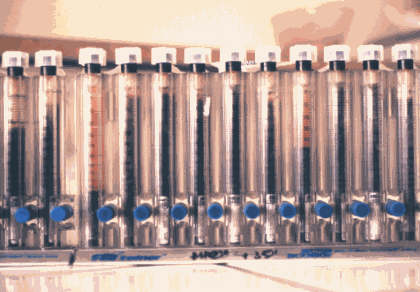
The patients also underwent a full blood count and liver function tests. The erythrocyte sedimentation rate was performed on a venous blood sample withdrawn directly into the vacutainer tube (Becton Dickinson), placed on a specific Seditainer carrier with a 2 mm scale (Figure 1).
|
|
|
Fig 1: Seditainer carrier (V.S.) |
CRP was measured by kinetic nephelometry on a Beckman Array instrument or on a Behring turbidimeter (Figure 2).
|
|
|
Fig 2: Behring turbidimeter (CRP) |

Both tests require approximately the same time, i.e. 1 hour.
Blood counts showed the patients' haemoglobin concentration to be than 11 g/l in 20 cases, a normal leukocyte count in 19 cases and thrombocytopaenia (<100.000 pl./mm3) in 21 cases. Liver function tests revealed raised transaminases in 20 cases. The average erythrocyte sedimentation rate of the patients was 19.8 mm on D1 and 39.2 mm on D7. In 5 cases, the erythrocyte sedimentation rate was greater than 105 mm.
|
Patients |
D1 |
D7 |
||
|
ESR |
CRP |
ESR |
CRP |
|
|
5 |
9 |
58 |
24 |
16 |
|
9 |
40 |
107 |
16 |
9 |
|
10 |
14 |
42 |
44 |
17 |
|
11 |
18 |
213 |
32 |
10 |
|
12 |
18 |
85 |
80 |
21 |
|
14 |
16 |
62 |
28 |
6 |
|
17 |
13 |
92 |
58 |
7 |
|
23 |
12 |
78 |
76 |
11 |
|
25 |
10 |
81 |
63 |
10 |
The mean CRP was 94.5 mg/l on D1 and 13.20 mg/l on D7. CRP was raised by D2 in all cases (Table I).
The increase in erythrocyte sedimentation rate is due to red blood cell agglutination and rouleaux formation due to the presence of positively charged inflammatory proteins in plasma, which neutralise the natural repulsive negative charges on red blood cells [1,2,3]. A large number of factors are involved in changing the erythrocyte sedimentation rate (Table II).
| RAISED ESR | DECREASED ESR |
|
Infectious diseases Anaemia Macrocytosis Hyperliproteinaemia Female sex (menstruation) Pregnancy (> 10th week) The elderly Obesity Oestrogen-progestagen therapy Heparin therapy Macromolecular solutions Chronic renal failure Gammopathies Severe injury
Causes of errors: |
Sickle Cell Anaemia Anisocytosis Spherocytosis Microcytosis Haemoglobinopathies Polycythaemia Greatly raised leukocytosis Hyperfibrinogenaemia Liver failure Cachexia High dose corticosteroids Acetyl salicylic acid Non-steroidal anti-inflammatory agents
Causes of errors: |
Table II: Factors changing the ESR
The normal ESR is 3 to 5 millimeters in men and 7 to 20 millimeters in women.
CRP is the most widely used of the acute phase inflammatory proteins because of its early rise and rapid kinetics [4]. It has a half life is 5 to 6 hours. CRP is concentrated in tissues involved in the inflammation where it exerts its biological properties: activation of complement, indirect bacteriostatic activity by facilitating ingestion of micro-organisms, facilitating resorption of damaged tissue by phagocytosis and activating platelet aggregation. In addition, CRP is believed to prevent entry of the sporozoite into the hepatocyte [5]. Normal CRP concentrations are 0 to 8 mg/l and do not differ according to sex or age [6]. CRP is raised in certain disorders (Table III).
| DISEASES | Raised CRP | CRP little or not raised |
|
Rheumatological |
Rheumatoid arthritis Horton's disease Ankylosing spondylitis Behcet's disease Vasculitides… |
Systemic lupus erythematosis Dermatomyositis Scleroderma Gougerot-Sjogren's Syndrome |
|
Gastro-intestinal |
Crohn's Disease Acute appendicitis Acute peritonitis Acute Pancreatitis |
Haemorrhagic Ulcerative colitis |
|
Malignancies |
Lymphomas Certain solid tumours |
Leukaemia |
|
Ischaemic |
Myocardial infarction | Coronary artery disease |
|
Traumatic |
Head injury with fracture Burns Surgery |
Uncomplicated head injury |
|
Infectious |
Bacterial infections: Pneumonia, meningitis, septicaemia Upper urogenital tract infections Acute prostatitis |
Viral infections: Lower urogenital tract infections Chronic prostatitis |
Table III: Change in the C reactive protein (CRP)
Mononuclear cells, which are activated by the Plasmodium during a malarial attack, produce inflammatory cytokines such as Tumor Necrosis Factor (TNF) interleukin 1 (IL1) or interleukin 6 (IL 6) [7, 8]. These cytokines stimulate the hepatic synthesis of acute phase inflammatory proteins including CRP [9], orosomucoid and haptoglobin, which all rise in malaria [10].
In contrast, a fall in haptoglobin reflects the presence of haemolysis [11]. More red blood cells are destroyed than are infected with the Plasmodium because of immune complexes which bind to the non-parasite infected erythrocytes and which are then destroyed by macrophages from the reticulo-histocyte system [8]. In addition, TNF inhibits erythropoiesis in the bone marrow. Haemolysis leads to the release of haemoglobin which binds to haptoglobin, reducing serum haptoglobin concentrations [12].
The rise in CRP in malaria has already been reported by other authors [13, 14]. In contrast to other authors, our study has not found any relationship with the severity of the parasitaemia [15]. CRP was greatly elevated (from 42 to 231 mg/l) in 17 cases. CRP has also been used as a good positive predictive indicator for the diagnosis of malaria in febrile people returning from a tropical area [13], and is a marker for malaria in epidemiological studies [16].
The rise in plasma immunoglobulin concentrations in patients suffering from malaria explains why the erythrocyte sedimentation rate increases. The sedimentation rate, however, remained normal in 50% of our patients [14]. Other studies have failed to find any significant differences between patients suffering from malaria and those suffering from other febrile conditions [17]. The rise in erythrocyte sedimentation rate is also influenced by anaemia, as was the case in 2 of our patients [1].
Twelve of the patients in our study had a normal sedimentation rate, whereas the CRP was raised in all of these cases. Dissociation between a normal sedimentation rate and raised CRP has already been reported for other inflammatory diseases [18], including those in children [19].
The changes in these two parameters were extremely interesting. The CRP fell significantly (p = 0.004) between day 1 (mean 95 mg/l) and day 7 (mean: 13 mg/), corroborating results from other studies on this subject [20]. The CRP increased from the first hours of inflammation and fell between D1 (mean: 20 mm) and D7 (mean 39 mm); difference not significant (p = 0.01). It required 3 to 6 weeks to return to normal.
In conclusion, the erythrocyte sedimentation rate appears to be inadequate to make a rapid diagnosis of the acute phase inflammatory reaction, whereas CRP offers excellent predictive value. In addition, the early rise in CRP as soon as the inflammatory reaction develops, makes it a distinctly preferable investigation to the sedimentation rate amongst the diagnostic factors used for malaria. It is, of course, the blood film which remains the fundamental investigation to confirm this diagnosis, allowing both the causative species and parasitaemia to be established. These are essential factors in the therapeutic management of the disease.
-
DUBOST J.J., SOUBRIER M., MEUNIER MN., SAUVEZIZ B. De la vitesse de sédimentation au profil inflammatoire. Rev. Méd. Int., 1994, 15, 727-733.
-
GENEREAU T., HERSON S. Vitesse de sédimentation augmentée, orientation diagnostique. Rev. Prat., 1993, 43, 241-245.
-
WENG X., CLOUTIER G., BEAULIEU R., GHISLAINE 0., ROEDERER G.O. Influence of acute-phase protein on erythrocyte aggregation. Am. Jour. Phys., 1996, 271, 346-352.
-
PAWLOTSKY Y., CHALES G., Etude des variations matinales de la sigma VS, de la vitesse de sédimentation (Westergreen) et de la protéine réactive C. Rev. Rhum., 1985, 52, 35-40.
-
NUSSLER A., PIED S., PONTET M., MILTGEN F., RENIA L., GENTILINI M., MAZIER D. Inflammatory status and preerythrocytic stage of malaria : role of the C reactive proteine. Exp. Parasit., 1991, 72, 1-7.
-
BOURÉE P., LANÇON A., RODRIGUE J.C. La protéine C réactive ou C.R.P. Tech. Biol., 1997, 3, 63-64.
-
GRANINGER W., THALHAMMER F., HOLLENSTEIN U., ZOTTER G.M., KREMSNER P.G. Serum protein concentration in Plasmodium falciparum malaria. Acta Trop., 1992, 52, 121-128.
-
WARREL D.A. Pathophysiologie du paludisme grave. Cahiers Santé., 1993, 3,276-279.
-
SUHRBIER A., ROYNES J.G., WALBY M.I., MC ADAM W.J., SINDEN R.E. C reactive protein and the stage of Plasmodium vivax and P. berghei. Trans. Roy. Soc. Trop. Med. Hyg., 1990, 84, 781.
-
GILLESPIE S.H., DOW C., RAYNES J.G., BEHRENS R.H., CHIODINI P.L., McADAM K.P.W.J. Measurement of acute phase proteins for assessing severity of Plasmodium falciparum malaria. Jour. Clin. Path., 1991, 44, 228231.
-
McGUIRE W., D'ALESSANDRO U., OLALEYE B.O., THOMSON M.C., LANGEROCK P., GREENWOOD B.M., KWIATOWSKI D. C reactive protein and haptoglobin in the evolution of a community-based malaria control programme. Trans. Roy. Soc. Trop. Med. Hyg., 1996, 90, 10-14.
-
ENGLER R. Protéines de la réaction inflammatoire. Fonctions régulatrices. Ann. Biol. Clin., 1988, 46, 336-342.
-
CHAGNON A., YAO N., CARLI P., PARIS J.F., MARLIER, PIERRE C., BUSSIERE H. La protéine C réactive dans l'accès palustre. Presse Méd., 1992, 21, 5, 217-218.
-
ERIKSSON B., HELLGREN U., ROMBO L., Changes in erythrocyte sedimentation rate C reactive protein and hematological parameters in patients with acute malaria. Scand. Jour. Inf. Dis., 1989, 21, 435-441.
-
NAIK P., VOLLER A. Serum C reactive protein levels and falciparum malaria. Trans. Roy. Trop. Med. Hyg. 1984, 78, 812-813.
-
HURT N., SMITH T., TANNER M., MWANKUSYE S., BORDMANN G., WEISS N.A., TEUSCHER T. Evolution of C reactive protein and haptoglobin as malaria episode markers in an area of high transmission in Africa. Trans. Roy. Soc. Trop. Med. 1994, 88, 182-186.
-
EGART T., GOSSET D., SAVINEL P., DEVULDER B., DELCAMBRE B. La protéine C réactive. Sem. Hôp. Paris., 1989, 65, 273-274.
-
HACHULA E. Vitesse de sédimentation ou protéine C réactive, que choisir ? N. P. N. Médecine., 1991, 173,133-136.
-
LELONG M., PARICHET D. VS ou CRP en pratique pédiatrique : vitesse de sédimentation ou mesure de la protéine C réactive ? N. P. N. Médecine., 1991, 171, 30-32.
-
NICOLAS P., HOVETTE P., MEROUZE F., TOUZE J.E., MARTER G. Cytokines et paludisme, étude du TNF alpha, de l'IL1 béta, de l'IL6 et du RIL2s chez 28 malades. Bull. Soc. Path. Exo., 1994, 87, 91-96.
| PDF file |
Home | Top of Page | Malaria 2000 | Previous Issue | Links | Congress | Contact Us